【Bilateral hip dysplasia】Biologic total hip replacement was successful
Breed: Border Collie, Weight: 24.8kg, Age: 8 years and 8 months, Sex: Male
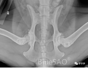
This is a middle aged male meteorite colored Border Collie. He has bilateral hip dysplasia, which has resulted in severe bilateral hip degenerative disease. He has bone spurs growing all over his bilateral hip joints, with the right side being far more severe than the left. The acetabulum on the left side still has a shape, while the right side is already a dish-shaped hip. The hind limbs trembled bilaterally when standing. But it is such a bad condition, still very active, and has to catch Frisbee.
Hip replacement surgery like this can be challenging and should not be rushed into by beginners. It occurs to me that we made such a presumptuous mistake years ago. At that time experience was far from adequate to deal with a similarly bad and complex condition. But for a beginner to encounter a case like this, the urge to try is certainly there. This is good, but it is also important to always maintain this passion to face and solve difficult conditions. It is also important to respect the objective laws and objectively assess your own strength. What kind of strength to fight what kind of monster. And it is very easy for a beginner to perform surgery on such a hip and to be discouraged if it fails.
Canine hip dysplasia is a genetic disorder and a developmental disorder. It is genetically related and it is not a congenital disorder. Congenital disorders are found at birth, such as harelip and cleft palate. Dogs with the hip dysplasia gene are born with normal hip joints. As the dog ages, the hip dysplasia phenotype gradually manifests itself. At a young age, the hip joint becomes lax. Because of the laxity, the femoral head cannot fully act on the acetabulum during stress. The acetabulum is not able to develop into a normal acetabulum that is deep enough to encase the femoral head. Because of the laxity, the femoral head tends to dislocate dorsally under normal stresses. The force on the acetabulum is then shifted from the normal crypt position to the dorsal side, resulting in a variety of wide and shallow acetabulums. Factors such as the nutritional status, activity level, and living environment of the affected dog itself can also influence the rate and severity of deterioration of hip dysplasia during the developmental process.
A very important and challenging aspect of total hip replacement for the dish-shaped hip in this case was to find the location of the original acetabulum. Only the new acetabulum polished from the original acetabulum can wrap around the artificial acetabular cup. How do you find it? From the development of hip dysplasia above, we know that the diseased acetabulum moves dorsally, so the original acetabulum must be located ventrally to the existing diseased acetabulum. So is there any anatomical landmark that allows us to determine intraoperatively that the polished one is the correct acetabulum? The answer is yes! Orthopaedic surgeons know that the acetabulum has a crypt, and that the crypt does not change with the position of the acetabulum. Therefore, finding the crypt during intraoperative polishing means finding the original acetabulum. Another issue is the size of the artificial acetabular cup. Since the diseased acetabulum is wide and shallow, the acetabular opening must be larger than the original acetabulum. Therefore, the preoperative measurement of the acetabulum must be larger than the actual acetabulum needed. The artificial acetabular cup chosen based on this data must also be larger. The new acetabulum will lose much of its anterior-posterior and dorsal edges, and the new acetabulum will not be able to wrap around the cup. Eventually it will fail. What to do? Choose an artificial acetabular cup (downsize) that is one size smaller than the measured one as the target.
After calibration according to the Marker in the iBlueVet operating system, the appropriate femoral stem prosthesis and acetabular cup prosthesis are selected. Preoperative planning was performed using a #5 biological femoral stem, a 24mm biological cup, and prepared +0, +3, and +6 16mm femoral head prostheses. Intraoperative testing was performed using a femoral head prosthesis test model before deciding on the final femoral head prosthesis.
Planedimplants: a #5 biological femoral stem, a 24mm biological cup, and prepared +0, +3, and +6 16mm femoral head prostheses.
preoperativex-rays
Log in to view the full medical record
Please Login to View AllOrthopedic Instruments We Use BlueSAO