A case of THR surgical treatment after femoral head resection in a two-year-old cat
Breed: A cat, Gender: male, Weight: 4.3 kg, Age: 2yearsold.
The cat started limping 6 months ago after falling from a building. Observation at home for a month, although the limp has improved, but has not fully recovered. The femoral neck fracture in the cat was found after examination at a hospital near the pet owner's home.The femoral head resection was subsequently performed on the cat. After the surgery, the cat still walks with obvious lameness. The muscles of the cat's affected limb were significantly atrophied. Although after half a year of rehabilitation and pain relief, there is still no obvious improvement. Instead, the cat becomes more and more afraid to walk on the affected limb.After the owner communicated with me, he brought the cat to our hospital for examination and found that the cat had obvious pain when the left hind limb was stretched. Subsequent x-rays showed extensive hyperplasia around the hip joint and femoral neck, as well as an indistinct acetabular fossa and significant thickening of the medial cortex. With such severe joint hyperplasia, the cat's quality of life must be seriously affected. The cat is also very resistant to being pulled on its hind limbs. I, therefore, recommend that the cat is treated with a total hip replacement (THR).
We first used BlueSAO iBlueVet orthopedic planning software to give this catan accurate preoperative planning. He was then given a new artificial joint using BlueSAO Cemented Hip Replacement Systemfor small dogs and cats.
The planned implants used were a #2 cemented femoral stem, a 12mm cemented cup, and an 8mm (+0) femoral head with 8mm (+2) and 8mm (+4) femoral head spares.
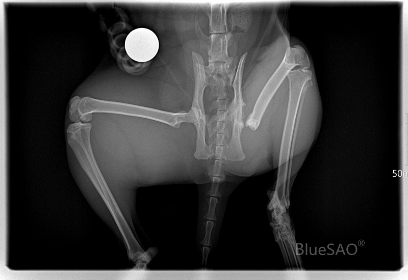
Preoperative X-rays
Four preoperative X-rays of the anteroposterior pelvis,lateral pelvis, anteroposterior femur, and lateral femur were taken.And precise planning is carried out using BlueSAO iBlueVet orthopedic planning software.
The X-ray showed that the muscles of the affected limb of this cat were severely atrophied, the shape of the acetabular fossa was not clear, a large number of osteophytes were formed, and the femoral neck was excessively resected. Moreover, the greater trochanter moved up significantly, which greatly increased the difficulty of cup placement, femoral medullary canal reaming, and postoperative reset.
Log in to view the full medical record
Please Login to View AllOrthopedic Instruments We Use BlueSAO