20201129 -【Comminuted fracture of middle tibia and fibula】A cat, fixed with PRCL-S system successfully
Clinical Syndromes and Medical history:
An unneutered male cat, about 6 months old, fell from the first floor and caused a comminuted fracture of the right middletibia and fibula.
Patient assessment:
This is a young cat with strong healing ability and a relatively quiet personality. The owner can cooperate well with the doctor.
Fracture assessment:
Closed comminuted fracture of therightmiddletibia and fibula. In terms of the degree of comminution and displacement, this is a high-energy fracture. There is less soft tissue coverage from the middle to the distal part of the tibia, which provides a less than ideal biological environment for fracture repair and may lead to delayedunionor nonunion of the fracture area. In addition to providing strong fixation, weshould also try to preserve the blood supply to the fracture area.
Repairing method options:
The type 2 external fixator can be considered, but postoperative care is relatively cumbersome. Because there are obvious short oblique bone fragments at the fracture site, Kirschner wires and wirecerclage are not recommended for fixation, and the cerclage wire willcause great damage to soft tissues. This is an irreducible comminuted fracture, so it is difficult for the wire to achieve 100% stability and the probability of failure is very large. Similarly, it is unlikely to achieve fracture reconstruction with lag screws. Minimally invasive techniques are the best choice, because the proximal and distal tibia bothhave enough spaceto place three screws. While the bone plate is used for fixation, the Intramedullary pin can improve the strength and rigidity of the overall structure. Therefore, for this case, we used BlueSAOPRCL-S6.5mm locking plate + intramedullarypin, and placed the implant through the MIPO technology, which not only provided stable fixation for the fracture, but also followed the BO principle.
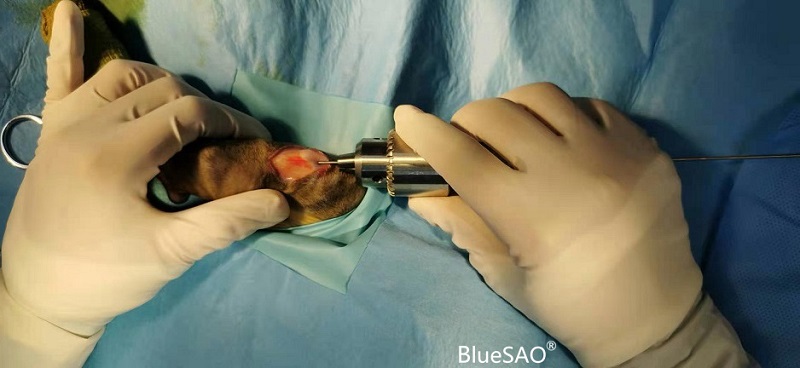
Operation report:
The cat wasplaced in a lateral position and an incision was maderespectivelyat the proximal and distal tibia. First,the intramedullary pin was placedalong thelong axisof the tibia.The knee jointwas bent, andthe intramedullary pin was inserted at the caudal side along the medial edge of the straight ligament of knee.The bones of young animals are relatively soft, so use a hand drill for needle insertion. After the intramedullary nailenters the proximal medullary cavity, it can enter the distal medullary cavity with the aid of a fluoroscopy assistant equipment. Subsequently, a periosteal dissector was used to bluntly peel a subcutaneous tunnel from the proximal to distal to facilitate the insertion of the bone plate. Then, the nearest and farthest screws were placed respectively, and the other screws were screwed in one by one, and the incision was routinely closed.
Postoperative evaluation of fracture:
Criteria | Assessment | Detail |
Alignment | good | Sagittal plane and frontal plane axially aligned well |
Bonding(fracture healing) | good | There is a large bone fragment on the lateral side of the fracture area, and the fracture gaps in other parts are very small, indicating that there is a complete periosteum. |
Material Choice | good | The size of the intramedullary pinis selected appropriately. However, the intramedullary pinhits the growth plate of the proximal tibial tubercle, which may cause avulsion fracture of the growth plate or affect the growth of the tibia. The size, lengthof the bone plate and the lengthof the screw are suitable. Both the proximal and distal screws did not affect the growth plate. |
Log in to view the full medical record
Please Login to View AllOrthopedic Instruments We Use BlueSAO